
| Connect & Subscribe |
Inside The Brain Of The REM Sleeper
Written by Carl DeGuzman, Spring 2010
Brain imaging technology has allowed us a glimpse into the fascinating world of what is happening inside our heads while we are in REM sleep. We can now see which parts of the brain are active during REM sleep and how that affects our sleep. Many of the findings explain the interesting phenomena that happen during REM sleep, particularly relating to the dreams that happen during this period.

Two parts of the region are of particular interest: the primary visual cortex and the extrastriate visual areas. Both are located in the back of the brain, and have to do with (you guessed it) vision-related things. The first region, the primary visual cortex, has the task of making sense of what we see in the outside world. It tells us what is happening around us by detecting patterns of light and interpreting them.
During REM sleep, it is mostly silent. This is not a big surprise, as most of the time while we sleep our eyes are obviously closed and cannot receive much visual stimuli. However, the extrastriate visual areas, which basically take the data from the primary visual cortex and tells us what is happening around us, are a lot more active in the brain than you would expect with no external stimuli. This can possibly help explain how we come up with such elaborate visual dreams, and along with other parts of the brain it helps to create a dream world complete with colors, textures, tastes, and smells.
Another part of the brain that shows intense activity during REM sleep is the limbic system, which is highly involved with emotions. Two areas of the limbic system in particular show a lot of activity, the hippocampus and the amygdala. The hippocampus is involved in turning short-term memories into long-term ones, which may help explain why many times we dream about things that were relevant to us recently. It also helps us in making associations, such as when we use a mnemonic device to memorize something. This feature may explain why we associate things that at first are seemingly unrelated, but if you look closer can in some way be explained. The amygdala on the other hand, is highly involved in fear and aggression. Obviously, this could explain the violent nature of some dreams, or one could even imagine an overactive amygdala leading to nightmares.
Interestingly, a part of the brain that is closely related to the limbic system, the frontal cortex, is relatively quiet during REM sleep. The frontal cortex, and in particular the prefrontal cortex, is involved in thought and judgment. In other words it helps reign in our animal instincts, as a professor of mine liked to say. This makes perfect sense, explaining why we sometimes dream of doing things that we know we would never do in real life, but seem perfectly normal while we are in the dream world.
Finally, what part of the brain is the trigger for REM sleep? Researchers have found that a region on the brainstem called the pons is necessary for REM sleep to happen. Various studies have shown that if you knock out part of one of the pons, known as the nucleus reticularis pontis oralis (or RPO for short), REM sleep cannot happen. Furthermore, two other nuclei in the pons that produce neurotransmitters related to keeping us awake must shut down before REM sleep can begin.
The brain stem is also the source of REM sleep paralysis, an important feature because if our body was as active as our brain during REM sleep, we would be able to act out our dreams which could lead to potentially harmful consequences. Unfortunately, this is actually possible, along with a number of other REM sleep disorders, which you can continue to read about here.
Carl used www.thebrain.mcgill.ca as a reference for this article.
 Subscribe To Our RSS Feed


About This Site
Welcome! This site is continuously being created by students of Dr. William C. Dement's Sleep And Dreams course at Stanford University.
We made this site as a call to action for people all over the world to live healthier, happier, safer, and more productive lives by learning about their own sleep. We have faith that reading the information provided on this site will motivate you to be smart about your sleep deprivation and strategic about your alertness in order to live life to your fullest, most energetic potential.
In fact, we challenge you to do so! What do you say, are you up for the challenge?
Interviews With Sleep Specialists: Insights Into the Worlds of Sleep Medicine & Sleep Business
America's Most Dangerous Disorder: What Is Sleep Apnea Doing To Your Sleep?
Sleep Debt: How Much More Will You Achieve When You Reduce Yours?
The Stages Of Sleep: The Journey Through The Night
Delayed Sleep Phase: You Want To Sleep But You're Not Tired Yet
Paralyzed at Night: Is Sleep Paralysis Normal?
Sleep In Words: Smart, Strange, and Funny Quotes About Sleep
Sleep Disorders In Children: What's Keeping Your Child From A Full Night's Rest?
Attacks of Pavor Nocturnus (a.k.a. Sleep Terrors, Night Terrors, or Incubus Attacks)
The Stanford Sleep Book

Dr. Dement's pioneering textbook has been the core text for Sleep and Dreams since 1980, but it has just recently been made available to the wider public for the first time.
In it you'll find a more detailed account of the most important things you need to know about sleep, alertness, dreams, and sleep disorders. Studies, statistics, plus plenty of Dr. Dement's classic anecdotes painting the history of sleep medicine.
Preface | Intro | Contents | Get A Copy
More Sleep Resources
The Zeo
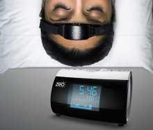
A revolution in personal sleep tracking, the Zeo is a wireless headband that transmits your brainwaves in realtime to a dock (pictured here) or your smartphone. The result? You can wake up and see exactly what stages of sleep you were in during the night! Unprecedented personalized sleep knowledge.
Sleep Paralysis: A Dreamer's Guide

Ever woken up paralyzed? A surprising number of us have, believe it or not. But few know the actual causes of this phenomenon, and fewer still how to exert control over it. Dream researcher and sleep paralysis expert Ryan Hurd shares breakthrough insights into how to do just that.
Important Disclaimer
Please Note:
The information found on this page and throughout this site is intended for general information purposes only. While it may prove useful and empowering, it is NOT intended as a substitute for the expertise and judgments of healthcare practitioners.
For more info, see our
Terms of Use.